Lymphocyte differentiation and detection of anti-enzyme antibody synthesis in lymph node cells after peroxidase (HRP) immunization
Wolf D. Kuhlmann
Laboratory Diagnostics & Cell Science, 56112 Lahnstein
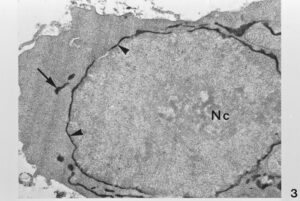
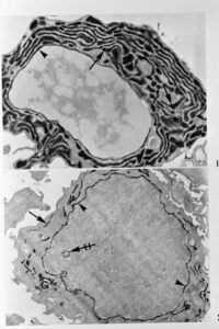
Mature plasma cell and lymphocytic blast cell synthesizing specific antibody in PNS and RER, the Golgi apparatus (G) is also stained. Note tangentially cut invaginations of PNS in the nucleus
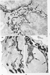
Plasma cells containing anti-HRP antibody in PNS, RER and transport of antibody toward the Golgi complex (G). Lower right side: an invagination of the positive PNS (arrow head) is cut tangentially, note nuclear pores (arrows)
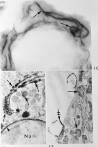
Blast cell as seen in the electron microscope of a 1 µm thick Epon section with interconnected sytem of antibody containing RER lamellae (arrows). Below, left side: Golgi complex of a plasma cell with antibodies in its lamellae and vesicles (arrows), the PNS (arrow head) is also stained. Below, right side: Plasma cell with antibody in RER cisternae (arrows). Note broken RER cistern at cell periphery


