AFP expression in hepatocytes and biliary epithelial cells (oval cells) during liver regeneration and chemical hepatocarcinogenesis
Wolf D. Kuhlmann
Laboratory Diagnostics & Cell Science, 56112 Lahnstein
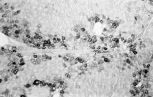
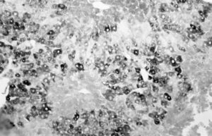
Rat liver tissue on days 2 to 4 after GalN injury with necrosis, inflammatory infiltrates, ductular proliferation and ballooning hepatocytes. Paraffin sections with AFP immunostaining or HE staining, and silver impregnation according to Gomori. AFP positive biliary epithelial cells (oval cells) often form tubular structures within portal tracts and periportal areas. Such histological features combined with cellular AFP immuno-expression occur quite similar in the case of liver repair after NNM intoxication. Magnification views 160 x, 250 x and 540 x
Rat liver from day 28 to 35 during high dose NNM feeding, some animals were pulse labelled with 3H thymidine. Paraffin sections with AFP immunostaining, some sections also with HE staining and autoradiography. Note AFP positive canalicular epithelial cells. Detection of AFP in gouped oval cells, also with ductular appearance forming tubular structures. Thymidine incorporation is observed in oval-shaped cells as well as in structures of ductular appearance. Magnification views 160 x, 250 x and 540 x
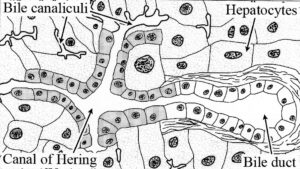
Structure of normal liver lobule with the canal of HERING which drains bile from the canaliculi into the bile duct (Junqueira L.C. & Carneiro [1980]; Basic Histology, Lange Medical Publica-tions p 350) with modifications observed in studies on oval cell proliferation during liver regeneration (KUHLMANN WD 1978, KUHLMANN WD and PESCHKE P 2006). Oval cells originate from the canals of HERING and are the source of AFP positive canalicular epithelial cell structures


